When assessing ophthalmic signs related to the adnexa, you’re evaluating the structures around the eye, not the eyeball itself. These include:
-
Eyelids
-
Lacrimal apparatus
-
Conjunctiva
-
Orbital tissues
This is crucial in both general eye exams and in identifying infections, trauma, tumors, or systemic diseases.
👁️🗨️ What Are the Ocular Adnexa?
| Adnexal Structure | Function |
|---|---|
| Eyelids | Protect the globe, distribute tear film |
| Lacrimal gland/system | Produces and drains tears |
| Conjunctiva | Mucous membrane lining eyelid and eyeball |
| Orbit & surrounding soft tissues | Protect and support the eye |
🔍 Ophthalmic Signs: Adnexa Exam Overview
1. Eyelids
Check for:
-
Ptosis – drooping of the eyelid (e.g., Horner’s syndrome, oculomotor palsy)
-
Lid retraction – seen in thyroid eye disease
-
Ectropion – lid turns outward
-
Entropion – lid turns inward (can cause lashes to scratch cornea)
-
Edema or swelling – common in infection or trauma
-
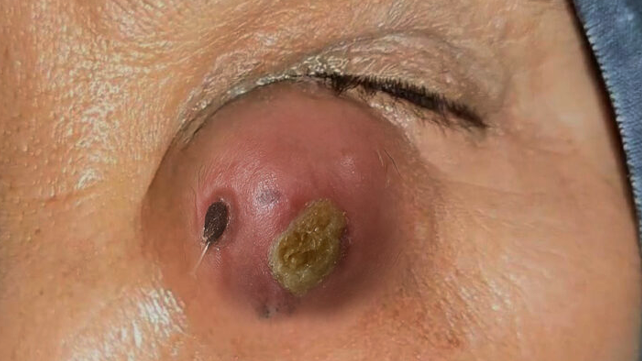
Lid masses – e.g., chalazion, hordeolum (stye), basal cell carcinoma
📌Signs:
Redness
Crusting
Incomplete closure (lagophthalmos)
Eyelid spasm (blepharospasm)
2. Lacrimal System
Inspect:
-
Lacrimal gland (upper outer lid) for swelling or tenderness (e.g., dacryoadenitis)
-
Tear drainage system for:
-
Epiphora – excessive tearing (obstruction)
-
Discharge or pus at puncta (suggests dacryocystitis)
-
Pressure on lacrimal sac – look for regurgitation of pus
-
3. Conjunctiva
Observe both:
-
Palpebral conjunctiva (lining eyelids)
-
Bulbar conjunctiva (covering sclera)
Look for:
-
Injection (redness) – general or localized
-
Chemosis – conjunctival edema (allergy, trauma, infection)
-
Pseudomembranes – suggest viral/bacterial conjunctivitis
-
Foreign bodies
-
Subconjunctival hemorrhage – bright red patch (benign or traumatic)
4. Orbital Signs
Observe for:
-
Proptosis – eye pushed forward (thyroid eye disease, tumor, orbital cellulitis)
-
Enophthalmos – eye sunken in (orbital fracture)
-
Periorbital swelling or bruising (trauma or infection)
-
Eye movement limitation – can indicate mass or muscle entrapment
-
Pain on movement – orbital cellulitis
📌 Check for Relative Afferent Pupillary Defect (RAPD) if optic nerve is involved.
5. Palpation and Special Tests
-
Gently palpate orbital rim for step-offs (trauma)
-
Palpate lacrimal sac for tenderness/discharge
-
Use fluorescein to detect foreign bodies or corneal abrasion
-
Test eyelid eversion to inspect upper conjunctiva
🧠 Clinical Conditions with Adnexal Signs
| Condition | Adnexal Signs |
|---|---|
| Orbital cellulitis | Proptosis, restricted movement, swollen red lids |
| Preseptal cellulitis | Lid swelling but no proptosis or movement restriction |
| Chalazion | Painless eyelid lump |
| Hordeolum (stye) | Painful red bump on eyelid |
| Basal cell carcinoma | Non-healing ulcer on lid |
| Thyroid eye disease | Lid retraction, proptosis, diplopia |
| Dacryocystitis | Swelling at medial canthus, purulent discharge |
| Conjunctivitis | Redness, discharge, chemosis |
🧪 Optional Diagnostic Tools
-
Slit lamp exam – for closer look at lids/conjunctiva
-
CT orbit – if abscess, trauma, or mass is suspected
-
Tear break-up time or Schirmer’s test – for dry eye
-
Culture – of discharge if infection suspected
✅ Summary: Key Signs in Adnexal Examination
| Sign | Meaning |
|---|---|
| Ptosis | CN III palsy or Horner’s syndrome |
| Proptosis | Thyroid orbitopathy, cellulitis, tumor |
| Chemosis | Allergy, infection |
| Epiphora | Nasolacrimal duct obstruction |
| Lid mass | Benign or malignant lesion |
| Eyelid retraction | Hyperthyroidism |
| Discharge from punctum | Dacryocystitis |
🩺 Treatment of Common Ocular Adnexal Conditions
1. Blepharitis
-
Management: Warm compresses (15 minutes, twice daily) to loosen crusts and massage the eyelids to express meibomian gland secretions.
-
Medications: Topical antibiotics like bacitracin or erythromycin ointment may be applied to the eyelid margin.
-
Hygiene: Regular eyelid scrubs with diluted baby shampoo or commercial lid cleansers to remove debris and bacteria.
2. Chalazion
-
Initial Treatment: Warm compresses (15 minutes, four times daily) to promote drainage.
-
If Persistent: Incision and drainage may be necessary if the chalazion persists after four weeks of medical therapy.
-
Steroid Injection: In some cases, a corticosteroid injection may be used to reduce inflammation.
3. Hordeolum (Stye)
-
Acute Management: Warm compresses (15 minutes, four times daily) to promote drainage.
-
Antibiotics: Topical antibiotics like bacitracin or erythromycin ointment may be applied.
-
If Non-Resolving: Incision and drainage may be required for persistent lesions.
4. Dacryocystitis
-
Acute Cases: Oral antibiotics (e.g., amoxicillin-clavulanate) to treat infection.
-
Chronic Cases: Surgical intervention, such as dacryocystorhinostomy, may be necessary to relieve obstruction.
5. Conjunctivitis
-
Bacterial: Topical antibiotics (e.g., ciprofloxacin 0.3% eye drops) are prescribed.
-
Viral: Typically self-limited; supportive care is provided.
-
Allergic: Antihistamine eye drops (e.g., olopatadine) and avoiding allergens.
6. Carcinoma in Situ of Ocular Adnexa
-
Surgical Excision: Primary treatment involves complete surgical removal of the neoplastic tissue.
-
Topical Chemotherapy: Agents like 5-fluorouracil or mitomycin C may be used for superficial lesions.
-
Photodynamic Therapy: Utilized for patients who are not candidates for surgery.
🎥 Educational Video: Ocular Adnexa Examination
For a comprehensive understanding and visual demonstration of ocular adnexal examination techniques, you can watch the following video:
This video provides step-by-step guidance on examining the eyelids, lacrimal apparatus, and surrounding structures, which is essential for diagnosing adnexal conditions.